Resolution: Spitzoid lesion on the leg
Diagnosis: spitzoid lesion, confirmed by histopathology. A 43-year-old woman with a new, growing lesion on the leg.
At first glance the lesion looks organized, but it fits no benign nevus pattern and shows no dermatofibroma criteria – so we move on to step 2A: no criteria for squamous cell carcinoma, but criteria for melanoma. So it had to come out.
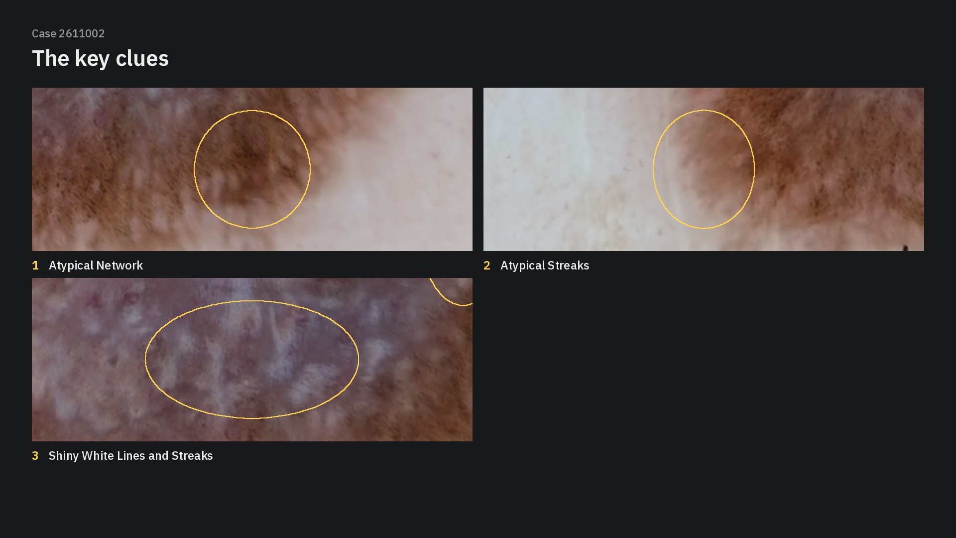
The three decisive clues:
Atypical streaks – irregular projections at the periphery
Atypical network – irregular lines and meshes
Shiny white lines – in the center, visible in polarized light
🎬 The case in a short video:
2611002_short_9x16.mp4Take-home: a new, growing spitzoid lesion in an adult with melanoma clues has to be excised – dermoscopy cannot separate a Spitz nevus from a spitzoid melanoma.
📊 Poll results – thanks to all 74 of you who voted!
Dermatofibroma – 34%
Clark (dysplastic) nevus – 3%
✅ Spitzoid melanocytic lesion – 8%
Invasive melanoma – 54%
Pigmented basal cell carcinoma – 1%
Only 8% chose the diagnosis confirmed by histopathology, a spitzoid lesion. More than half of you (54%) went for invasive melanoma – an understandable call, because the atypical streaks, atypical network and shiny white lines are melanoma clues, and dermoscopy cannot separate a Spitz nevus from a spitzoid melanoma. Another 34% voted dermatofibroma, likely because of the white lines in the center – but the lesion shows no dermatofibroma criteria, and with melanoma clues in a new, growing lesion the management is the same either way: excision.
💬 Thanks Blandine for pointing straight at the white lines and the edge of the lesion – exactly where the decisive clues were.
Which clue convinced you first? Share it in the comments.
Evidence Club members: the full case solution with the algorithm walk-through, every criterion explained and this week's teaching point is ready → Case Solution. Not a member yet? See the Dermoscopy Evidence Club → learn.dermoscopy.ch/dermoscopy-evidence-club
Case courtesy of Julien Anriot.