Resolution: Invasive melanoma on the back
Diagnosis: invasive melanoma, confirmed by histopathology; Breslow thickness 0.4 mm. A 67-year-old man; the lesion on the back was found at a routine skin examination.
The lesion is disorganized, so we go straight to step 2A – and melanoma criteria are present. Nine were documented; three stand out:
Atypical network – irregular lines and meshes
Negative pigment network – pale lines around darker areas
Shiny white lines – visible in polarized light
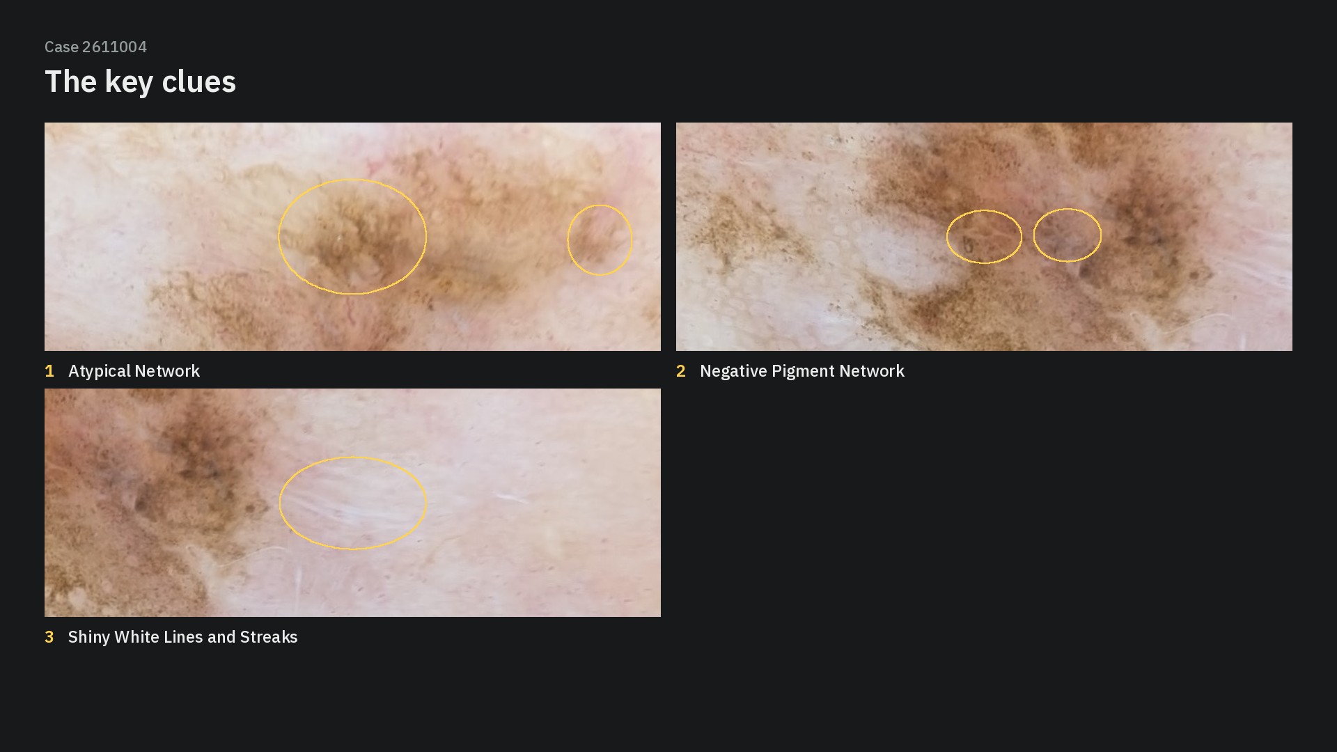
Take-home: even a thin melanoma found at a routine examination can show the full spectrum of melanoma criteria – it is the combination that makes the diagnosis.
📊 Poll results – thanks to all 60 of you who voted!
Clark (dysplastic) nevus – 3%
Melanoma in situ – 37%
Seborrheic keratosis – 5%
Invasive melanoma ✅ – 47%
Pigmented basal cell carcinoma – 8%
Almost half of you got it exactly right with invasive melanoma, and another 37% chose melanoma in situ – so 84% recognized a melanoma. The split between the two is understandable: with a Breslow thickness of only 0.4 mm this is a thin melanoma, and deciding between in situ and invasive was the hardest call in this case. As the take-home says, it is the combination of criteria that makes the diagnosis.
💬 Thanks Rus for your detailed step-by-step walk-through of the algorithm in the comments!
Which clue convinced you first? Share it in the comments.
Evidence Club members: the full case solution with the algorithm walk-through, every criterion explained and this week's teaching point is ready → Case Solution. Not a member yet? See the Dermoscopy Evidence Club → learn.dermoscopy.ch/dermoscopy-evidence-club