Resolution: Dermatofibroma on the lower leg
Diagnosis: dermatofibroma, confirmed by histopathology. A 47-year-old man with a slowly growing lesion on the lower leg.
The lesion is disorganized, so we go straight to step 2A: no criteria for basal cell carcinoma, none for squamous cell carcinoma, but criteria for melanoma – so it had to come out.
The three decisive clues:
Shiny white lines – visible only in polarized light
Atypical network
Milky-red area
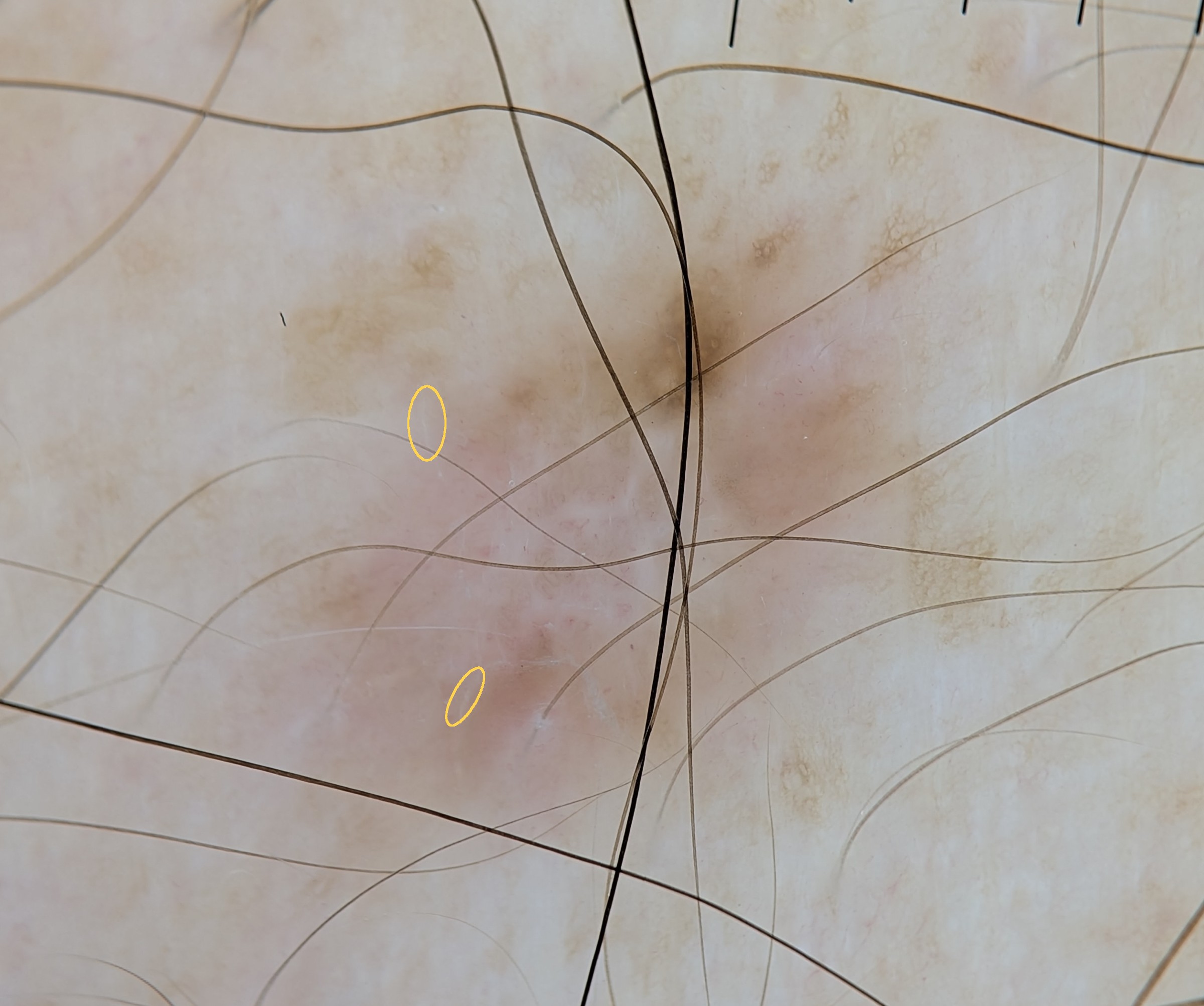
Take-home: shiny white lines, dotted vessels and milky-red areas also occur in dermatofibromas; in a disorganized lesion with melanoma clues the algorithm still says excise, and histopathology decides.
📊 Poll results – thanks to all 33 of you who voted!
Dermatofibroma: 40% ✅
Invasive melanoma: 36%
Dysplastic nevus: 15%
Basal cell carcinoma: 9%
Spitz nevus: 0%
So 40% of you got the diagnosis right, but almost as many chose melanoma. That split is the whole point of this case: shiny white lines, an atypical network and a milky-red area occur in both. In a disorganized lesion with melanoma clues, the algorithm says excise, and only histopathology settles it.
Which clue convinced you first? Share it in the comments.
Evidence Club members: the full case solution with the algorithm walk-through, every criterion explained and this week's teaching point is ready → Case Solution. Not a member yet? See the Dermoscopy Evidence Club → learn.dermoscopy.ch/dermoscopy-evidence-club
Want to see the whole spectrum? Universe of Dermatofibromas is the complete lecture on this lesion – included in All Access, 20% off for Evidence Club members → Universe of Dermatofibromas